
Skin Cancer Diagnosis
You Could Be At Risk Of Skin Cancer
It only takes one irregular mole to become a Melanoma.
Two in three Australians will be diagnosed with Skin Cancer by the time they are 70.
Melanoma is the third most common cancer in Australia in both women and men.
A Skin Check Is Easy
The earlier a skin cancer is detected and treated the better your chance of avoiding surgery and potential disfigurement.
At the clinic, we perform a full body skin check, and our Doctor performs a full review and assessment. This involves mapping every mole or mark on your body.
Get Familiar With Your Skin & Body
You should familiarise yourself with your skin, and be aware of any changes that might suggest a skin cancer.
Look for anything changing uniquely on your skin anywhere for more than 2-3 weeks.
Crusty sores, things that don't heal, new spots, freckles, or moles, or other things (lesions) that change in colour or thickness.
If you notice any of these changes, arrange an appointment with our Doctors for a skin check immediately.
The good news is that skin cancer is one of the most preventable forms of cancer in Australia. Having regular skin checks will help you be aware of any skin changes.
The Skin Check Process
At our clinic, we employ a new level in skin cancer checks using advanced technology to ensure your diagnosis and ongoing treatment are of the highest standard.
Through every stage:
- Initial skin check
- Any treatments, to
- Follow-ups or Annual Checks
The team aim to offer the best possible information, treatment and care.
Your journey starts with our "5 Step Skin Assessment Process"
Our 5 Step Method Ensures Accuracy
- Consult - Listen and Take a Complete Clinical History
- Scan - Comprehensive Full Body Scan
- Skin Map - systemic analysis locates all pigmented skin-changes in fine detail including moles, sunspots, hyper- and hypo-pigmentation, nodules, ulcers, dry skin and more
- Collect - 12 ultra high definition images using our computer controlled process for maximum reproducibility.
- Dermoscopic Focus - on lesions at risk of change or areas of special interest in full 4K brilliance.
- Record - specific sunspot characteristics are stored in a patient database with all important imagery and data including:
- Dermoscopy or microscopic images in full 4K brilliance,
- Body Mapped or macroscopic imagery
- Overview location images
- Evaluate - Each individual spots’ key metrics employing ABCD Analysis - Asymmetry, Borders and Areas, Colours and Variability, Diameter
- Diagnose - All images are reliably, quickly and immediately assessed by both a Doctor and computer-based modelling. Borderline lesions are classified by their dermoscopic features by combining three internationally accepted analytic methods:
- ABCD Risk Factors
- MacKenzie Analysis
- An integrated expert system using neural network comparisons to calculate and classify each spot with a value. These values are based on the world’s largest multi-centre skin cancer study DANAOS.
- Assess Risk Factors - our Doctor personally reviews images in the context of all relevant patient history data
- Report - In combination with your Doctor’s review detailed images help inform the patients of their
- skin status,
- diagnosis and
- the course of action
Expert Diagnosis of Suspicious Moles
Accuracy is important when dealing with melanoma, as you don’t get a second chance.
Our expert Skin Cancer Doctors are among the world’s most experienced and have diagnosed over 80,000 patients.
Skin Cancer Education
Knowledge is power, which is why we want you to know what lesions to look out for and how to protect you and your family. Many patients have identified skin cancer on their friends and family members after their Mole Mapping appointment with us.
Head-to-toe Skin Check
Our skilled Melanographers, advanced technology and experience Doctors will conduct a thorough examination of your skin, assessing moles using a dermatoscope – a device used to view the intricate structure of a mole. Moles that meet our criteria will be imaged using a specialised digital dermoscopic camera to create a view of the internal structure (dermoscopic image) and the external structure (clinical image) for subsequent medical diagnosis and comparison.
Follow Up Skin Check Process
The Clinic helps patients stay safe with efficient and accurate follow-up examinations.
At follow up appointments the scanning is repeated and any new skin damage or pigmented skin-changes can be immediately recognised.
- Comparisons - Each new image captured at follow-up examinations is automatically positioned alongside previous images of the same lesion for direct comparison
- Changes - Details can be magnified up to 200x and by comparing earlier findings both you and your Doctor can see changes in the skin to fractions of a millimetre
- Expert System - Automatically calculates any changes and offers deep insights that can assist in early diagnosis.
- Skin Management - Clinical imagery document pre- and post-operative treatment status
- Report - In combination with your Doctor’s review detailed images help inform the patients of:
- Any sun-damage or skin aging
- Current skin status and Diagnosis and
- Your best course of action
Over time a rich sequential history of your sunspots is built allowing changes in your skin to be immediately identified and analysed leading to the earliest intervention of any known method.
Your Skin Type
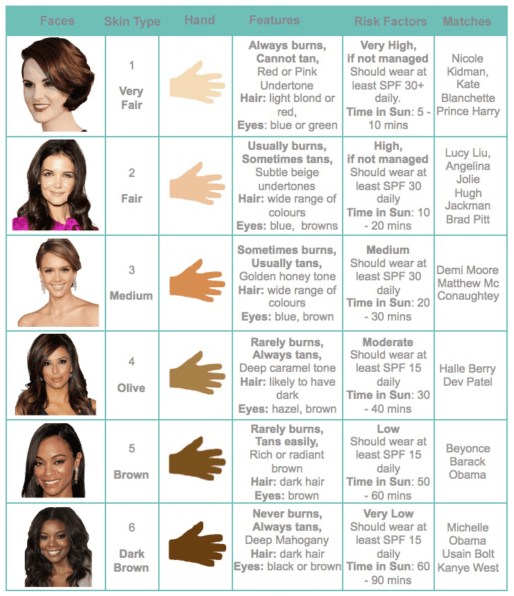
EVERYONE BENEFITS FROM REGULAR SKIN CHECKS
This should include checking areas that are normally hidden from the sun, because skin cancer can develop anywhere on the body.
Show our Team anything you find that is new or different on your skin, especially if it is changing uniquely in any way for more than 2 to 3 weeks.